Login to your account

Username *
Password *
Remember Me

Blog With Right Sidebar

A microRNA gene signature for identification of lung cancer.

Related Articles

Lung cancer is the leading cause of cancer deaths worldwide, compounded by late diagnosis. MicroRNAs (miRNA) are recently discovered short, noncoding genes that play essential roles in tissue differentiation during normal development and tumorigenesis. miRNA profiles across all histologic grades can provide a reliable and standardized method for the identification of lung cancer.

METHODS: A microRNA lung cancer dataset was analyzed. Differentially expressed microRNAs were obtained post-normalization of data using t-test (p < 0.01). The data for differentially expressed microRNAs were processed using K-nearest neighbors classification method to obtain unique miRNAs expression patterns. The predicted mRNA targets were identified using TargetScan and the molecular functions associated with the predicted targets were retrieved from the Gene Ontology Consortium and represented using GO IDs in a directed acyclic graph.

RESULTS: The results indicate that lung cancer samples can be classified using a small panel of 19 unique microRNAs (8 down-regulated and 11 up-regulated) with over 85% classification accuracy. Furthermore, using classical enrichment analysis, this study identified 66 molecular function groups which are potentially the functional signaling pathways altered by these differentially expressed microRNAs.

CONCLUSIONS: We identified a microRNA gene signature representative of functioning as a diagnostic biomarker for lung cancer. These findings can potentially form the basis for the development of a standardized diagnostic assay that can be used for early diagnosis of lung cancer equally well from resection specimens and cytology samples.

Pericytes in Chronic Lung Disease.

Related Articles

pericytes
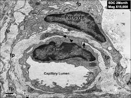
Pericytes are mesenchymal cells embedded within the abluminal surface of the endothelium of microvessels such as capillaries, pre-capillary arterioles, post-capillary and collecting venules, where they maintain microvascular homeostasis and participate in angiogenesis.

In addition to their roles in supporting the vasculature and facilitating leukocyte extravasation, pericytes have been recently investigated as a subpopulation of mesenchymal stem cells (MSCs) due to their capacity to differentiate into numerous cell types including the classic MSC triad, i.e. osteocytes, chondrocytes and adipocytes.

Other studies in models of fibrotic inflammatory disease of the lung have demonstrated a vital role of pericytes in myofibroblast activation, collagen deposition and microvascular remodelling, which are hallmark features of chronic lung diseases such as asthma, chronic obstructive pulmonary disorder, pulmonary fibrosis and pulmonary hypertension.

Further studies into the mechanisms of the pericyte-to-myofibroblast transition and migration to fibrotic foci will hopefully clarify the role of these cells in chronic lung disease and confirm the importance of pericytes in human fibrotic pulmonary disease. © 2014 S. Karger AG, Basel.


Usefulness of Lung Ultrasound in the Diagnosis of Community-acquired Pneumonia in Children.

Related Articles

EchoPneumonia is a life-threatening disease in children. With the current lack of universal diagnostic criteria, the diagnosis is usually made on clinical manifestations and findings from chest radiographs. Ultrasonography has recently been applied to the detection of pulmonary diseases. However, few data have been published showing its effectiveness in detecting pneumonia in children. The objective of this study was to determine the power of lung ultrasonography (LUS) for the diagnosis of pneumonia in children.

MATERIALS AND METHODS: This retrospective study was carried out by reviewing medical records. Patients admitted to a pediatric ward with a diagnosis of pneumonia from June 1, 2010 to December 31, 2012 were enrolled in this study. Personal information, laboratory data, characteristics on LUS scan, and the results of chest radiography and LUS were collected. We compared the detection rate of pneumonia by chest radiography and LUS. LUS scans were followed up in 23 patients during the progression of their disease.

RESULTS: A total of 163 patients was enrolled. Chest radiography was able to detect pneumonia in 152 patients, whereas LUS detected pneumonia in 159 patients. In LUS, the positive rates of the comet-tail sign, air bronchograms, fluid bronchograms, vascular pattern within the consolidation, and pleural effusion were 50.9%, 93.7%, 20.1%, and 28.9%, respectively. During follow up, the average size of the pneumonia patch in 23 patients decreased from 10.9 ± 8.7 cm(2) to 5.5 ± 8.2 cm(2), and finally to 2 ± 1.9 cm(2) on Day 1, Days 3-5 and Days 7-14, respectively.

CONCLUSION: LUS is a sensitive diagnostic tool with which to identify pneumonia in children. It is also useful in following up the progress of pneumonia. We suggest that LUS is a complementary tool to chest radiography in the diagnosis of pneumonia in children and that the follow up of pneumonia by LUS can reduce the exposure of children to ionizing radiation.

High resolution computed tomography and classification of children with interstitial lung diseases.

High resolution computed tomography (HRCT) was used to classify children with interstitial lung diseases (ILD).

Sixty children with ILD underwent HRCT in supine position under free-respiratory conditions during scanning. Children under 5 years old were sedated with chloral hydrate and the scanning scope was from the lung apex to the diaphragm. In children older than 5 years old, scans were obtained at three levels: aortic arch, tracheal carina, and 1 cm above the right diaphragm. Five infectious patients were followed up. Two experienced radiologists read the films blindly to observe the type and distribution of ground-glass opacities and bronchovascular bundle abnormalities.

Bronchovascular bundles were thick in 49 patients, and were thick and stiff in 27 patients. Of the 41 infectious patients, 39 showed thickened bronchovascular bundles, and 26 showed thick and stiff bronchovascular bundles. Of the 19 non-infectious patients, bronchovascular bundles were thickened in 10 patients, and were thick and stiff in 1 patient. Forty-one patients showed lobular ground-glass opacity (32 infectious, 9 non-infectious). Twenty-seven patients showed both bronchovascular bundle abnormality and lobular ground-glass opacity (20 infectious, 7 non-infectious). Eighteen patients showed patchy or mosaic ground-glass opacity (16 infectious, 2 non-infectious). There were 4 cases of bronchiectasis.

HRCT is the first non-invasive diagnostic method for children with ILD, and its different manifestations can be classified. In early manifestation, bronchovascular bundles were abnormal and complicated with lobular ground-glass opacity. Patchy ground-glass opacity was the most common manifestation, and appeared to be difficult to disappear. Bronchiectasis indicated that the disease is irretrievable.

Therapeutic approach to respiratory infections in lung transplantation.

Related Articles

Lung transplant recipients (LTRs) are at life-long risk for infections and disseminated diseases owing to their immunocompromised state. Besides organ failure and sepsis, infection can trigger acute and chronic graft rejection which increases mortality. Medical prophylaxis and treatment are based on comprehensive diagnostic work-up including previous history of infection and airway colonisation to reduce long-term complications and mortality.

Common bacterial pathogens include Pseudomonas and Staphylococcus, whilst Aspergillus and Cytomegalovirus (CMV) are respectively the commonest fungal and viral pathogens. Clinical symptoms can be various in lung transplant recipients presenting an asymptomatic to severe progress. Regular control of infection parameters, daily lung function testing and lifelong follow-up in a specialist transplant centre are mandatory for early detection of bacterial, viral and fungal infections. After transplantation each patient receives intensive training with rules of conduct concerning preventive behaviour and to recognize early signs of post transplant complications.

Early detection of infection and complications are important goals to reduce major complications after lung transplantation.

Search