Login to your account

Username *
Password *
Remember Me

Blog With Right Sidebar

Imaging of community-acquired pneumonia: Roles of imaging examinations, imaging diagnosis of specific pathogens and discrimination from noninfectious diseases.

Related Articles

This article reviews roles of imaging examinations in the management of community-acquired pneumonia (CAP), imaging diagnosis of specific CAP and discrimination between CAP and noninfectious diseases.

Chest radiography is usually enough to confirm the diagnosis of CAP, whereas computed tomography is required to suggest specific pathogens and to discriminate from noninfectious diseases. Mycoplasma pneumoniae pneumonia, tuberculosis, Pneumocystis jirovecii pneumonia and some cases of viral pneumonia sometimes show specific imaging findings. Peribronchial nodules, especially tree-in-bud appearance, are fairly specific for infection. Evidences of organization, such as concavity of the opacities, traction bronchiectasis, visualization of air bronchograms over the entire length of the bronchi, or mild parenchymal distortion are suggestive of organizing pneumonia.

We will introduce tips to effectively make use of imaging examinations in the management of CAP.

The IASLC/ATS/ERS classification of lung adenocarcinoma-a surgical point of view.

Related Articles

adenocarcinomaoflung40x01
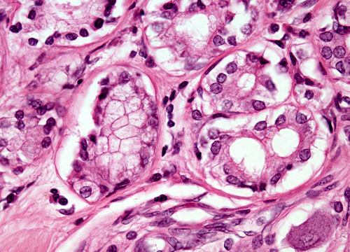
Adenocarcinoma has become the most common histologic type of lung cancers. Ground glass nodules (GGN), most of them early stage noninvasive or minimally invasive adenocarcinomas (MIA), have been encountered more frequently with the application of computed tomography (CT) screening.

The International Association for the Study of Lung Cancer (IASLC)/American Thoracic Society (ATS)/European Respiratory Society (ERS) histologic lung adenocarcinoma classification combines radiologic, histologic, clinic, and molecular features to form a diagnostic approach for different subgroups of diseases. One of the major focuses of this new classification is the introduction of adenocarcinoma in situ (AIS) and MIA, to replace the old term of bronchioloalveolar carcinoma (BAC). Not all GGNs are malignant lesions that should be surgically resected upon first presentation. A management approach different to solid nodules has been suggested based on the understanding that these lesions tend to have a more indolent nature. Hasty intervention should be avoided and potential surgical risks, radiation exposure, patient psychology, and socio-economical burden must be balanced comprehensively before surgery is decided upon. In the mean time, surgical issues concerning extent of resection and lymphadenectomy should also be carefully contemplated once intervention is deemed necessary. Extremely good prognosis with a near 100% disease-free survival could be expected when a pure GGN is completely resected. This has led to re-evaluation of sublobar resections, including both segmentectomy and big wedge resection, for small (≤2 cm) less invasive histology (AIS or MIA) appearing as GGN on CT scan. Evidences are accumulating that these limited resections are oncologically equivalent to standard lobectomy. And extensive lymph node dissection may not have additional staging or prognostic benefit. These would add new meaning to the contemporary definition of minimally invasive surgery for lung cancers.

Overall, joint effort from a multiple disciplinary team is imperative, and decision making should be based on both anatomical and biological nature of the disease.

Inhaled tyrosine kinase inhibitors for pulmonary hypertension: a possible future treatment.

Related Articles

Inhaled tyrosine kinase inhibitors for pulmonary hypertension: a possible future treatment.

Drug Des Devel Ther. 2014;8:1753-63

Authors: Pitsiou G, Zarogoulidis P, Petridis D, Kioumis I, Lampaki S, Organtzis J, Porpodis K, Papaiwannou A, Tsiouda T, Hohenforst-Schmidt W, Kakolyris S, Syrigos K, Huang H, Li Q, Turner JF, Zarogoulidis K

Abstract
Pulmonary hypertension is a disease with severe consequences for the human body. There are several diseases and situations that induce pulmonary hypertension and are usually underdiagnosed. Treatments include conventional medical therapies and oral, inhaled, intravenous, and subcutaneous options. Depending on its severity, heart or lung transplant may also be an option. A possible novel treatment could be tyrosine kinase inhibitors. We conducted experiments with three jet nebulizers and three ultrasound nebulizers with erlotinib, gefitinib, and imatinib. Different residual cup designs and residual cup loadings were used in order to identify the best combination to produce droplets of less than 5 μm in mass median aerodynamic diameter. We found that gefitinib could not be transformed into a powder, so conversion to an aerosol form was not possible. Our experiments indicated that imatinib is superior to erlotinib with regard to small droplet size formation using both inhaled technologies (1.37 μm <2.23 μm and 1.92 μm <3.11 μm, jet and ultrasound, respectively) and, at jet devices (1.37 μm <1.92 μm). Cup designs C and G contribute best to small droplet creation uniquely supporting and equally well the activity of both drugs. The disadvantage of the large droplets formed for erlotinib was offset when combined with residual cup C (1.37 μm instead of 2.23 μm). At a 2 mL dose, the facemask and cone mouthpieces performed best and evenly; the facemask and low dose were the best choice (2.08 μm and 2.12 μm, respectively). Erlotinib and imatinib can be administered as an aerosols, and further in vivo experimentation is necessary to investigate the positive effects of these drugs in the treatment of pulmonary hypertension.

PMID: 25336919 [PubMed - in process]

Pneumothorax: from definition to diagnosis and treatment.

Related Articles

Pneumothorax: from definition to diagnosis and treatment.

J Thorac Dis. 2014 Oct;6(Suppl 4):S372-6

Authors: Zarogoulidis P, Kioumis I, Pitsiou G, Porpodis K, Lampaki S, Papaiwannou A, Katsikogiannis N, Zaric B, Branislav P, Secen N, Dryllis G, Machairiotis N, Rapti A, Zarogoulidis K

Abstract
Pneumothorax is an urgent situation that has to be treated immediately upon diagnosis. Pneumothorax is divided to primary and secondary. A primary pneumothorax is considered the one that occurs without an apparent cause and in the absence of significant lung disease. On the other hand secondary pneumothorax occurs in the presence of existing lung pathology. There is the case where an amount of air in the chest increases markedly and a one-way valve is formed leading to a tension pneumothorax. Unless reversed by effective treatment, this situation can progress and cause death. Pneumothorax can be caused by physical trauma to the chest or as a complication of medical or surgical intervention (biopsy). Symptoms typically include chest pain and shortness of breath. Diagnosis of a pneumothorax requires a chest X-ray or computed tomography (CT) scan. Small spontaneous pneumothoraces typically resolve without treatment and require only monitoring. In our current special issue we will present the definition, diagnosis and treatment of pneumothorax from different experts in the field, different countries and present different methods of treatment.

PMID: 25337391 [PubMed]

Pneumothorax after transbronchial needle biopsy.

Related Articles

Pneumothorax after transbronchial needle biopsy.

J Thorac Dis. 2014 Oct;6(Suppl 4):S427-34

Authors: Boskovic T, Stojanovic M, Stanic J, Pena Karan S, Vujasinovic G, Dragisic D, Zarogoulidis K, Kougioumtzi I, Dryllis G, Kioumis I, Pitsiou G, Machairiotis N, Katsikogiannis N, Papaiwannou A, Madesis A, Diplaris K, Karaiskos T, Zaric B, Branislav P, Zarogoulidis P

Abstract
Currently there several diagnostic techniques that re used by radiologists and pulmonary physicians for lung cancer diagnostics. In several cases pneumothorax (PNTX) is induced and immediate action is needed. Both radiologists and pulmonary physicians can insert a chest tube for symptom relief. However; only pulmonary physicians and thoracic surgeons can provide a permanent solution for the patient. The final solution would be for a patient to undergo surgery for a final solution. In our current work we will provide all those diagnostic cases where PNTX is induced and treatment from the point of view of expert radiologists and pulmonary physicians.

PMID: 25337399 [PubMed]

Search