Login to your account

Username *
Password *
Remember Me

Blog With Right Sidebar

Novel Aerosol Delivery Devices.

Delivery of medication to sites of action through airways has been used for centuries but has gained momentum in recent decades. Currently available modes of aerosol delivery offer advantages but still there are shortcomings. Delivery of active agents to sites of action is affected by many factors beyond the characteristics of the delivery devices, including the coordination between inhalation and actuation and dependence on the patient's inspiratory flow and breathing pattern.

Recent advances in drug delivery focus around changes in the generation of particle size with better penetration to the targeted sites, easier activation of the device with inspiratory flow, ease of use including fewer steps in using the device, and better portability. Availability of computer chips allows for smart delivery devices to tailor delivery to the patient's breathing pattern and lung function. Further, smart devices can provide feedback to patients.

Novel inhaler technologies, separately or in combination with new therapeutic agents, are in development not only for respiratory diseases but also for diseases of other systems. This article reviews some of the recent clinically relevant advances in aerosol delivery devices.

Stereotactic body radiotherapy for second pulmonary nodules after operation for an initial lung cancer.

pic1
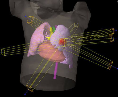
A second lung cancer is occasionally observed in patients who underwent surgical resection of the index lung cancer. The purpose of this study is to evaluate stereotactic body radiation therapy for second lung cancer.

METHODS: Fifty-one medically inoperable patients who underwent stereotactic body radiation therapy for second lung cancer were the subjects: 31 cases of multiple primary lung cancer and 20 of pulmonary metastasis from the index cancer. Clinical stage was T1a in 27 patients, T1b in 13 patients and T2a in 11 patients, and 70% of subjects had impaired respiratory function. Histology of second lung cancer was adenocarcinoma in 16 patients, squamous cell carcinoma in 9 patients and not assessed in 25 patients. The interval between index cancer operation and stereotactic body radiation therapy was 31 months (range: 4-171). The total stereotactic body radiation therapy doses were 48 Gy in 4 fractions or 60 Gy in 10 fractions.

RESULTS: With the median follow-up of 36 months, 3-year overall survival rates were 62% with the median survival time of 46 months. Cause-specific survival was 73% at 3 years. Overall survival for multiple primary lung cancer and pulmonary metastasis was quite similar: 62 and 61% at 3 years, respectively. Three-year overall survival was 77% for T1a and 43% for T1b or T2a. Grade 2 pulmonary toxicities occurred in five patients and one patient died of Grade 5 pneumonitis.

CONCLUSIONS: Even though the subjects were medically inoperable, the survival outcomes of stereotactic body radiation therapy were favorable. Furthermore, having acceptable toxicity, stereotactic body radiation therapy is feasible and could be an option for multiple primary lung cancer and pulmonary metastasis after surgical resection for the index cancer.

Bronchoscopy in patients with haemoptysis and normal computed tomography of the chest is unlikely to result in significant findings.

INTRODUCTION: The standard evaluation of haemoptysis in a department of respiratory medicine would currently consist of chest radiography, contrast-enhanced computed tomography (CT) and fibre-optic bronchoscopy (FOB), regardless of the result of the CT. Our aim was to evaluate whether patients presenting with haemoptysis but no positive finding on a contrast-enhanced CT of the chest are at risk for having serious disease, first of all lung cancer, and thus whether FOB is mandatory for such patients.

METHODS: We searched the literature and retrospectively reviewed all records of patients referred with haemoptysis between 2000 and 2010 at the Department of Respiratory Diseases and Allergy, Aarhus University Hospital, Denmark.

RESULTS: A total of 379 patient records were reviewed for inclusion in the clinical part of the study. Of these, 269 had the information required for the study and had been examined with CT. In all, 16 of the 269 patients were diagnosed with lung cancer. In all of these, a tumour or other findings indicating a possible tumour were seen on the chest CT. No additional cases of lung cancer were discovered during FOB, and no cases had been missed by the CT.

CONCLUSION: CT should be used as first-line examination in patients with a history of haemoptysis. Furthermore, in patients above 40 years of age with positive findings of any kind on the CT, further examination with FOB is indicated. However, if the chest CT is without pathological findings, it is most unlikely that FOB will reveal anything of significance.

FUNDING: none.
TRIAL REGISTRATION: not relevant.

A 3-cm Single-port Video-assisted Thoracoscopic Lobectomy for Lung Cancer.

Related Articles

thVideo-assisted thoracoscopic surgery is advantageous over traditional surgical practices, because of a faster postoperative recovery period, less pain, and a shorter hospital length of stay. There is no single standard technique in the video-assisted thoracoscopic surgery approach. Although these "minimally invasive" resections are habitually performed through 3-port or 4-port incision, we performed a left lower lobectomy in a 54-year-old male patient for lung cancer, through a 3-cm single-port incision.

Pulmonary Endogenous Fluorescence Allows the Distinction of Primary Lung Cancer from the Perilesional Lung Parenchyma.

Pre-therapeutic pathological diagnosis is a crucial step of the management of pulmonary nodules suspected of being non small cell lung cancer (NSCLC), especially in the frame of currently implemented lung cancer screening programs in high-risk patients. Based on a human ex vivo model, we hypothesized that an embedded device measuring endogenous fluorescence would be able to distinguish pulmonary malignant lesions from the perilesional lung tissue.

METHODS: Consecutive patients who underwent surgical resection of pulmonary lesions were included in this prospective and observational study over an 8-month period. Measurements were performed back table on surgical specimens in the operative room, both on suspicious lesions and the perilesional healthy parenchyma. Endogenous fluorescence signal was characterized according to three criteria: maximal intensity (Imax), wavelength, and shape of the signal (missing, stable, instable, photobleaching).

RESULTS: Ninety-six patients with 111 suspicious lesions were included. Final pathological diagnoses were: primary lung cancers (n = 60), lung metastases of extra-thoracic malignancies (n = 27) and non-tumoral lesions (n = 24). Mean Imax was significantly higher in NSCLC targeted lesions when compared to the perilesional lung parenchyma (p<0,0001) or non-tumoral lesions (p<0,0001). Similarly, photobleaching was more frequently found in NSCLC than in perilesional lung (p<0,0001), or in non-tumoral lesions (p<0,001). Respective associated wavelengths were not statistically different between perilesional lung and either primary lung cancers or non-tumoral lesions. Considering lung metastases, both mean Imax and wavelength of the targeted lesions were not different from those of the perilesional lung tissue. In contrast, photobleaching was significantly more frequently observed in the targeted lesions than in the perilesional lung (p≤0,01).

CONCLUSION: Our results demonstrate that endogenous fluorescence applied to the diagnosis of lung nodules allows distinguishing NSCLC from the surrounding healthy parenchyma and from non-tumoral lesions. Inconclusive results were found for lung metastases due to the heterogeneity of this population.

Search