Login to your account

Username *
Password *
Remember Me

Blog With Right Sidebar

Long-term non-invasive ventilation therapies in children: a scoping review protocol.

Related Articles

Non-invasive ventilation (NIV) in children has become an increasingly common modality of breathing support where pressure support is delivered through a mask interface or less commonly through other non-invasive interfaces. At this time, NIV is considered a first-line option for ventilatory support of chronic respiratory insufficiency associated with a range of respiratory and sleep disorders. Previous reviews on the effectiveness, complications and adherence to NIV treatment have lacked systematic methods. The purpose of this scoping review is to provide an overview of the evidence for the use of long-term NIV in children.

METHODS AND ANALYSIS: We will use previously established scoping methodology. Ten electronic databases will be searched to identify studies in children using NIV for longer than 3 months outside an intensive care setting. Grey literature search will include conference proceedings, thesis and dissertations, unpublished trials, reports from regulatory agencies and manufacturers. Two reviewers will independently screen titles and abstracts for inclusion, followed by full-text screening of potentially relevant articles to determine final inclusion. Data synthesis will be performed at three levels: (1) an analysis of the number, publication type, publication year, and country of publication of the studies; (2) a summary of the study designs, outcomes measures used; (3) a thematic analysis of included studies by subgroups.

ETHICS AND DISSEMINATION: This study will provide a wide and rigorous overview of the evidence on the use of long-term NIV in children and provide critical information for healthcare professionals and policymakers to better care for this group of children. We will disseminate our findings through conference proceedings and publications, and evaluate the results for further systematic reviews and meta-analyses.

Bronchoscopic lung volume reduction by endobronchial valve in advanced emphysema: the first Asian report.

Related Articles

Endobronchial valve (EBV) therapy is increasingly being seen as a therapeutic option for advanced emphysema, but its clinical utility in Asian populations, who may have different phenotypes to other ethnic populations, has not been assessed.

PATIENTS AND METHODS: This prospective open-label single-arm clinical trial examined the clinical efficacy and the safety of EBV in 43 consecutive patients (mean age 68.4±7.5, forced expiratory volume in 1 second [FEV1] 24.5%±10.7% predicted, residual volume 208.7%±47.9% predicted) with severe emphysema with complete fissure and no collateral ventilation in a tertiary referral hospital in Korea.

RESULTS: Compared to baseline, the patients exhibited significant improvements 6 months after EBV therapy in terms of FEV1 (from 0.68±0.26 L to 0.92±0.40 L; P<0.001), 6-minute walk distance (from 233.5±114.8 m to 299.6±87.5 m; P=0.012), modified Medical Research Council dyspnea scale (from 3.7±0.6 to 2.4±1.2; P<0.001), and St George's Respiratory Questionnaire (from 65.59±13.07 to 53.76±11.40; P=0.028). Nine patients (20.9%) had a tuberculosis scar, but these scars did not affect target lobe volume reduction or pneumothorax frequency. Thirteen patients had adverse events, ten (23.3%) developed pneumothorax, which included one death due to tension pneumothorax.

CONCLUSION: EBV therapy was as effective and safe in Korean patients as it has been shown to be in Western countries. (

TRIAL REGISTRATION: ClinicalTrials.gov: NCT01869205).

Prolongation of antibiotic treatment for cystic fibrosis pulmonary exacerbations.

Related Articles

Pulmonary exacerbations frequently lead to an irrevocable loss of lung function in cystic fibrosis (CF) patients. Although extended antibiotic duration has not been shown to be associated with improved outcomes in CF overall, it is not known whether there is a subset of patients who may benefit from longer treatment courses.

METHODS: This was a retrospective cohort study, using the Toronto CF Database from 1997 to 2012, of CF individuals with pulmonary exacerbations requiring intravenous antibiotic treatment. We investigated factors associated with improvement in forced expiratory volume in 1second (FEV1) in patients treated with ≤14days and >14days of antibiotic treatment.

RESULTS: A total of 538 pulmonary exacerbations in 253 patients were used for these analysis; 39% of these exacerbations fully recovered lung function at follow-up. Exacerbations were more frequently treated with >14days of antibiotics in older patients with lower FEV1 at exacerbation and higher rates of B. cepacia complex infections. Subjects with exacerbations treated for >14days had a significantly greater increase in FEV1 from day 14 to follow up compared to those with ≤14days (p<0.001). On multivariable analysis, smaller changes from days 0 to 14 of antibiotics and treatment duration>14days were associated with greater increases in FEV1 from day 14 to follow-up. In those who received >14days of antibiotic therapy, smaller improvements in FEV1 change from day 0 to 14 and younger age at exacerbation were significantly associated with a greater FEV1 response from day 14 to end of treatment. Antibiotic treatment >14days was not associated with longer time to subsequent exacerbation.

CONCLUSIONS: This study highlights that in the treatment of pulmonary exacerbations, maximum lung function is not achieved within 14days in all patients, and that there is continued improvement beyond this period.

Mosaic Attenuation: Etiology, Methods of Differentiation, and Pitfalls.

Mosaic attenuation is a commonly encountered pattern on computed tomography that is defined as heterogeneous areas of differing lung attenuation. This heterogeneous pattern of attenuation is the result of diverse causes that include diseases of the small airways, pulmonary vasculature, alveoli, and interstitium, alone or in combination.

Small airways disease can be a primary disorder, such as respiratory bronchiolitis or constrictive bronchiolitis, or be part of parenchymal lung disease, such as hypersensitivity pneumonitis, or large airways disease, such as bronchiectasis and asthma. Vascular causes resulting in mosaic attenuation are typically chronic thromboembolic pulmonary hypertension, which is characterized by organizing thrombi in the elastic pulmonary arteries, or pulmonary arterial hypertension, a heterogeneous group of diseases affecting the distal pulmonary arterioles.

Diffuse ground-glass opacity can result in a mosaic pattern related to a number of processes in acute (eg, infection, pulmonary edema), subacute (eg, organizing pneumonia), or chronic (eg, fibrotic diseases) settings. Imaging clues that can assist the radiologist in pinpointing a diagnosis include evidence of large airway involvement, cardiovascular abnormalities, septal thickening, signs of fibrosis, and demonstration of airtrapping at expiratory imaging. (©)RSNA, 2015.

Close Surveillance with Long-term Follow-up of Subjects with Pre-invasive Endobronchial Lesions.

cancer
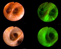
Autofluorescence bronchoscopy (AFB) and computed tomography (CT) enable lung cancer (LC) detection at early (pre-)invasive stage. However, LC risk in individuals harboring pre-invasive endobronchial lesions is unclear. OBJECTIVES To assess LC incidence and identify potential risk determinants in individuals harboring pre-invasive lesions.

METHODS In our tertiary care referral center, 164 individuals with pre-invasive lesions were monitored up to 12.5 years by repeated AFB and CT. Occurrence of LC was monitored. Clinical management depended on histological grade, with cancer patients receiving standard of care. Potential risk determinants (smoking status, baseline histology, cancer history and COPD-status) were evaluated in relation to cancer occurrence, event-free survival (EFS) and overall survival (OS). MEASUREMENTS AND MAIN

RESULTS During surveillance (median of 30 months, range 4-152) of 164 individuals with pre-invasive lesions (80 high-grade and 84 low-grade at inclusion), 61 LCs were detected in 55 individuals (median time-to-event 16.5 months). Twenty-three LCs (38%) were detected by CT, thirty-eight (62%) by AFB. More cancers (36/61;59%) developed from separate, rather than initial lesional sites. Individuals with high-grade lesions were more likely diagnosed with LC at the same or another site in the lungs than those with low-grade lesions (p=0.03). Independent risk determinants for OS were previous curatively treated cancer and COPD (p≤0.05).

CONCLUSIONS Presence of pre-invasive lesions, especially high-grade lesions, may serve as LC risk marker. LCs occur both at pre-invasive lesion sites and elsewhere in the bronchial epithelium or lung parenchyma. Prospective validation of biomarkers and randomized intervention studies are required to determine optimal management strategies.

Search