Login to your account

Username *
Password *
Remember Me

Blog With Right Sidebar

Pneumococcal Vaccine and Patients with Pulmonary Diseases.

Related Articles

Chronic pulmonary diseases describe chronic diseases that affect the airways and lung parenchyma. Examples of common chronic pulmonary diseases include asthma, bronchiectasis, chronic obstructive lung disease, lung fibrosis, sarcoidosis, pulmonary hypertension and cor pulmonale.

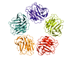
Pulmonary infection is considered a significant cause of mortality in patients with chronic pulmonary diseases. Streptococcus pneumoniae is the leading isolated bacteria from adult patients with community-acquired pneumonia, the most common pulmonary infection. Vaccination against S. pneumoniae can reduce the risk of mortality especially from more serious infections in both immunocompetent and immunocompromised patients.

Patients with chronic pulmonary diseases who take steroids or immunomodulating therapy (e.g., methotrexate, anti-TNF inhibitors), having concurrent sickle cell disease or other hemoglobinopathies, primary immunodeficiency disorders, human immunodeficiency virus infection / acquired immunodeficiency syndrome (HIV/AIDS), nephrotic syndrome, and hematologic or solid malignancies should be vaccinated with both 13-valent pneumococcal conjugate vaccine (PCV13) and the pneumococcal polysaccharide vaccine 23-valent (PPSV23).

Percutaneous Ultrasonography as Imaging Modality and Sampling Guide for Pulmonologists.

Related Articles

USonoG

Ultrasound (US) imaging is gradually progressing into common practice in contemporary pulmonology. Its main applications are to determine the presence and amount of pleural effusions and to guide subsequent treatment interventions. Guidelines recommend the use of US for these indications.

Training programs are organized and competency levels are formulated. Image guidance with US to obtain specimens for pathologic and/or microbiological analysis is less extensively practiced by pulmonologists but it is an important tool for tumour staging and diagnosing diseases. Lung tumours in contact with the pleural surface, pleural thickenings, mediastinal tumours and chest wall tumours are conceivable indications for pulmonologists to approach with the help of US visualization. Moreover, sampling of chest disease-related extrathoracal lesions may also be regarded as the working field of the pulmonologist. For example, supraclavicular and axillar lymph node metastasis, and also soft tissue and bone metastases, are lesions encountered during dissemination tests.

US-guided biopsy provides not only a diagnosis, but also gives information on the stage of disease in sometimes inaccessible primary lesions. US-guided sampling increases diagnostic efficacy and safety and enables very precise performance of fine-needle aspirations as well as tissue core biopsies. © 2014 S. Karger AG, Basel.

Immunoglobulin g expression in lung cancer and its effects on metastasis.

Related Articles

Lung cancer is one of the leading malignancies worldwide, but the regulatory mechanism of its growth and metastasis is still poorly understood.

We investigated the possible expression of immunoglobulin G (IgG) genes in squamous cell carcinomas and adenocarcinomas of the lung and related cancer cell lines. Abundant mRNA of IgG and essential enzymes for IgG synthesis, recombination activation genes 1, 2 (RAG1, 2) and activation-induced cytidine deaminase (AID) were detected in the cancer cells but not in adjacent normal lung tissue or normal lung epithelial cell line.

The extents of IgG expression in 86 lung cancers were found to associate with clinical stage, pathological grade and lymph node metastasis. We found that knockdown of IgG with siRNA resulted in decreases of cellular proliferation, migration and attachment for cultured lung cancer cells. Metastasis-associated gene 1 (MTA1) appeared to be co-expressed with IgG in lung cancer cells. Statistical analysis showed that the rate of IgG expression was significantly correlated to that of MTA1 and to lymph node metastases. Inhibition of MTA1 gene expression with siRNA also led to decreases of cellular migration and attachment for cultured lung cancer cells.

These evidences suggested that inhibition of cancer migration and attachment induced by IgG down-regulation might be achieved through MTA1 regulatory pathway. Our findings suggest that lung cancer-produced IgG is likely to play an important role in cancer growth and metastasis with significant clinical implications.

C-reactive protein and lung diseases.

Related Articles

CRP

C-reactive protein (CRP), a member of the pentraxin family of plasma proteins, is one of the most distinctive acute phase reactants. In response to inflammation, cell damage or tissue injury, plasma level of CRP rapidly and dramatically increases up to 1000-fold, a phenomenon that has been used for years to monitor infections and many destructive/inflammatory conditions.

The magnitude of CRP increase usually correlates with the severity of injury or inflammation and reflects an important physiological role of this interesting but still under-investigated protein. It is now generally accepted that CRP is involved in host defense and inflammation. However, the exact function of this protein in health and disease remains unclear. Many studies have demonstrated that in different pathophysiological conditions CRP might be involved in the regulation of lung function and may participate in the pathogenesis of various pulmonary disorders.

The fluctuation of CRP concentrations in both alveolar fluid and serum associated with different pulmonary diseases suggests its important role in lung biology. Discussion of the still controversial functions of CRP in lung physiology and diseases is the main focus of this review.

Metabolic activity measured by FDG PET predicts pathological response in locally advanced superior sulcus NSCLC.

Related Articles

PETSCAN

Pathological complete response and tumor regression to less than 10% vital tumor cells after induction chemoradiotherapy have been shown to be prognostically important in non-small cell lung cancer (NSCLC). Predictive imaging biomarkers could help treatment decision-making. The purpose of this study was to assess whether postinduction changes in tumor FDG uptake could predict pathological response and to evaluate the correlation between residual vital tumor cells and post-induction FDG uptake.

METHODS: NSCLC patients with sulcus superior tumor (SST), planned for trimodality therapy, routinely undergo FDG PET/CT scans before and after induction chemoradiotherapy in our institute. Metabolic end-points based on standardized uptake values (SUV) were calculated, including SUVmax (maximum SUV), SUVTTL (tumor-to-liver ratio), SUVpeak (SUV within 1 cc sphere with highest activity), and SUVPTL (peak-to-liver ratio). Pathology specimens were assessed for residual vital tumor cell percentages and scored as no (grade 3), <10% (grade 2b) and >10% vital tumor cells (grade 2a/1).

RESULTS: 19 and 23 patients were evaluated for (1) metabolic change and (2) postinduction PET-pathology correlation, respectively. Changes in all parameters were predictive for grade 2b/3 response. ΔSUVTTL and ΔSUVPTL were also predictive for grade 3 response. Remaining vital tumor cells correlated with post-induction SUVpeak (R=0.55; P=0.007) and postinduction SUVPTL (R=0.59; P=0.004). Postinduction SUVPTL could predict both grades 3 and 2b/3 response.

CONCLUSION: In NSCLC patients treated with chemoradiotherapy, changes in SUVmax, SUVTTL, SUVpeak, and SUVPTL were predictive for pathological response (grade 2b/3 and for SUVTTL and SUVPTL grade 3 as well). Postinduction SUVPTL correlated with residual tumor cells.

Search