Login to your account

Username *
Password *
Remember Me

Blog With Right Sidebar

Bronchoscopic Treatment of Emphysema: State of the Art.

Related Articles

In recent years, different bronchoscopic techniques have been proposed for the treatment of emphysema, with the aim of obtaining the same clinical and functional advantages of lung volume reduction surgical techniques while reducing risks and costs. Such techniques can be classified into: methods employing devices that block the airways (e.g. spigots and unidirectional valves),

methods that have a direct effect on the lung parenchyma (polymeric lung volume reduction, coils and thermal vapor ablation) and procedures that facilitate the expiration of trapped air from the emphysematous lung (airway bypass).

This review aimed to evaluate the indications, outcomes and safety of the different techniques, based on the evidence from the available literature. Results obtained by these methods are encouraging, but they are still based mainly on studies with small groups of patients. However, several trials are ongoing and in the near future we will acquire more knowledge which should lead to a better optimization of these procedures.

Meanwhile, the bronchoscopic treatment of emphysema cannot yet be considered a standard of care and patients should be treated in the context of clinical trials or controlled registries, with well-defined programs of evaluation and follow-up.

Temporary Endobronchial Embolization with Silicone Spigots for Moderate Hemoptysis: A Retrospective Study.

Related Articles


bleeding
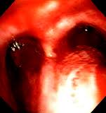
Background: The management of airway bleeding is generally performed in an emergency to prevent hypoxemia and lung flooding. When the bleeding arises from peripheral lesions that are not visible endoscopically, bronchoscopic options have limited curative intents. Endobronchial embolization using silicone spigots (EESS) is a novel approach. Objectives: We analyzed the efficacy and safety of EESS in a retrospective study.

Methods: We retrospectively reviewed charts of patients referred to our center for moderate hemoptysis (MH) who underwent EESS. Successful management is defined as immediate bleeding cessation.

Results: From December 2008 to January 2012, 9 patients were treated with EESS in our endoscopy unit. The MH originated from the left upper lobe in 4 cases, the right upper lobe in 3 cases and the right middle lobe and left lower lobe in 1 case each. Thirteen spigots were inserted. The success rate was 78%. Of the 9 patients, 7 were referred to interventional radiology for bronchial artery embolization, with a success rate of 86%, and 2 were referred for thoracic surgery. One patient had EESS as definitive treatment; the silicone spigots were bronchoscopically removed after a median of 4 days in 6 of the remaining 8 patients. Only 2 patients had hemoptysis recurrence after a median follow-up of 107 days (ranging from 13 to 1,017 days). None of the patients died from hemoptysis.

Conclusion: EESS is an original, temporary technique that requires only a flexible bronchoscope and biopsy forceps for placement and removal. EESS ensures airway protection while waiting for definitive management.

Standards for Quality Care in Respiratory Rehabilitation in Patients With Chronic Pulmonary Disease.

Related Articles

Respiratory rehabilitation (RR) has been shown to be effective with a high level of evidence in terms of improving symptoms, exertion capacity and health-related quality of life (HRQL) in patients with COPD and in some patients with diseases other than COPD. According to international guidelines, RR is basically indicated in all patients with chronic respiratory symptoms, and the type of program offered depends on the symptoms themselves.

As requested by the Spanish Society of Pneumology and Thoracic Surgery (SEPAR), we have created this document with the aim to unify the criteria for quality care in RR. The document is organized into sections: indications for RR, evaluation of candidates, program components, characteristics of RR programs and the role of the administration in the implementation of RR.

In each section, we have distinguished 5 large disease groups: COPD, chronic respiratory diseases other than COPD with limiting dyspnea, hypersecretory diseases, neuromuscular diseases with respiratory symptoms and patients who are candidates for thoracic surgery for lung resection.

The tuberculous granuloma: an unsuccessful host defence mechanism providing a safety shelter for the bacteria?

Related Articles

The tuberculous granuloma: an unsuccessful host defence mechanism providing a safety shelter for the bacteria?

Clin Dev Immunol. 2012;2012:139127

Authors: Silva Miranda M, Breiman A, Allain S, Deknuydt F, Altare F

Abstract
One of the main features of the immune response to M. Tuberculosis is the formation of an organized structure called granuloma. It consists mainly in the recruitment at the infectious stage of macrophages, highly differentiated cells such as multinucleated giant cells, epithelioid cells and Foamy cells, all these cells being surrounded by a rim of lymphocytes. Although in the first instance the granuloma acts to constrain the infection, some bacilli can actually survive inside these structures for a long time in a dormant state. For some reasons, which are still unclear, the bacilli will reactivate in 10% of the latently infected individuals, escape the granuloma and spread throughout the body, thus giving rise to clinical disease, and are finally disseminated throughout the environment. In this review we examine the process leading to the formation of the granulomatous structures and the different cell types that have been shown to be part of this inflammatory reaction. We also discuss the different in vivo and in vitro models available to study this fascinating immune structure.

PMID: 22811737 [PubMed - in process]

Sarcoidosis and tuberculosis cytokine profiles: indistinguishable in bronchoalveolar lavage but different in blood.

Related Articles

Sarcoidosis and tuberculosis cytokine profiles: indistinguishable in bronchoalveolar lavage but different in blood.

PLoS One. 2012;7(7):e38083

Authors: Thillai M, Eberhardt C, Lewin AM, Potiphar L, Hingley-Wilson S, Sridhar S, Macintyre J, Kon OM, Wickremasinghe M, Wells A, Weeks ME, Mitchell D, Lalvani A

Abstract
BACKGROUND: The clinical, radiological and pathological similarities between sarcoidosis and tuberculosis can make disease differentiation challenging. A complicating factor is that some cases of sarcoidosis may be initiated by mycobacteria. We hypothesised that immunological profiling might provide insight into a possible relationship between the diseases or allow us to distinguish between them.
METHODS: We analysed bronchoalveolar lavage (BAL) fluid in sarcoidosis (n = 18), tuberculosis (n = 12) and healthy volunteers (n = 16). We further investigated serum samples in the same groups; sarcoidosis (n = 40), tuberculosis (n = 15) and healthy volunteers (n = 40). A cross-sectional analysis of multiple cytokine profiles was performed and data used to discriminate between samples.
RESULTS: We found that BAL profiles were indistinguishable between both diseases and significantly different from healthy volunteers. In sera, tuberculosis patients had significantly lower levels of the Th2 cytokine interleukin-4 (IL-4) than those with sarcoidosis (p = 0.004). Additional serum differences allowed us to create a linear regression model for disease differentiation (within-sample accuracy 91%, cross-validation accuracy 73%).
CONCLUSIONS: These data warrant replication in independent cohorts to further develop and validate a serum cytokine signature that may be able to distinguish sarcoidosis from tuberculosis. Systemic Th2 cytokine differences between sarcoidosis and tuberculosis may also underly different disease outcomes to similar respiratory stimuli.

PMID: 22815689 [PubMed - in process]

Search